
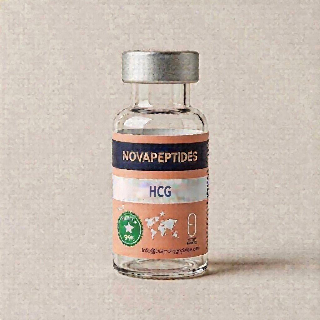
HCG (Human Chorionic Gonadotropin): The Glycoprotein Hormone for Reproductive Research


Price range: $85.00 through $165.00
HCG (Human Chorionic Gonadotropin) is a heterodimeric glycoprotein hormone fundamental to human reproduction and endocrine function. Produced by the syncytiotrophoblast during pregnancy, HCG maintains the corpus luteum and stimulates progesterone production essential for early gestation. Structurally composed of an alpha subunit identical to LH, FSH, and TSH, and a unique beta subunit conferring biological specificity, HCG binds to the luteinizing hormone/chorionic gonadotropin receptor (LHCGR) to regulate gonadal function. Beyond pregnancy, HCG serves critical clinical applications in fertility medicine, male hypogonadism treatment, and testosterone restoration while preserving spermatogenesis.
HCG: The Glycoprotein Hormone at the Intersection of Reproduction and Endocrinology
HCG represents one of the most clinically significant glycoprotein hormones in human biology, serving as the molecular signal that initiates and sustains early pregnancy while maintaining the delicate hormonal balance required for reproductive success. Unlike simple peptide hormones, HCG is a complex heterodimeric glycoprotein with sophisticated structural features that enable precise biological targeting. For researchers studying reproduction, endocrinology, and metabolic regulation, understanding HCG‘s structure and function provides essential insights into the broader family of glycoprotein hormones and their receptor interactions.
Molecular Structure and Glycoprotein Architecture
The molecular complexity of HCG distinguishes it from simpler peptide hormones. With a total molecular weight of approximately 36.7 kDa, HCG consists of two non-covalently associated subunits—alpha (α) and beta (β)—each with distinct structural and functional contributions.
The alpha subunit (~14.5 kDa) is structurally identical across all glycoprotein hormones including luteinizing hormone (LH), follicle-stimulating hormone (FSH), and thyroid-stimulating hormone (TSH). This shared subunit contains two N-linked glycosylation sites and is encoded by a single gene (CGA). Research published in Wikipedia and International Journal of Molecular Sciences confirms that this structural conservation enables the alpha subunit to participate in receptor binding and signal transduction across the glycoprotein hormone family.
The beta subunit (~22.2 kDa) confers biological specificity to HCG. While sharing 80-85% homology with the beta subunit of LH, the HCG beta subunit contains unique structural elements including an extended C-terminal tail with additional O-glycosylation sites. These differences explain why HCG and LH, despite activating the same receptor, exhibit distinct pharmacokinetic properties and biological half-lives.
Biological Production and Physiological Functions
In pregnancy, HCG is synthesized by the syncytiotrophoblast—the outer layer of the blastocyst that invades the uterine wall during implantation. Production begins shortly after fertilization, making HCG the earliest detectable marker of pregnancy. The hormone doubles approximately every 48-72 hours during the first trimester, reaching peak concentrations around weeks 8-11 before gradually declining as the placenta assumes progesterone production.
The primary physiological function of HCG is the rescue and maintenance of the corpus luteum—the ovarian structure that produces progesterone following ovulation. By binding to LH receptors on corpus luteum cells, HCG stimulates continued progesterone synthesis, preventing menstruation and creating a supportive environment for embryonic development. Without this HCG-mediated signal, the corpus luteum would degenerate, progesterone would fall, and pregnancy would terminate.
Beyond corpus luteum maintenance, HCG influences multiple pregnancy-related processes including:
- Immune Modulation: Protecting the fetal allograft from maternal immune rejection
- Angiogenesis: Promoting blood vessel development in the placenta
- Fetal Testicular Development: Stimulating testosterone production in male fetuses to support sexual differentiation
- Thyroid Function: Weak stimulation of the TSH receptor, contributing to gestational thyroid changes
Receptor Mechanism and Signal Transduction
HCG exerts its biological effects through the luteinizing hormone/chorionic gonadotropin receptor (LHCGR or LHR)—a G protein-coupled receptor expressed primarily on gonadal tissues. This receptor, as documented in ScienceDirect, binds both LH and HCG, explaining their overlapping biological activities.
Upon HCG binding, the LHCGR activates Gs alpha subunits, stimulating adenylate cyclase to increase intracellular cAMP levels. This second messenger activates protein kinase A (PKA), leading to phosphorylation of transcription factors and enzymes that mediate steroidogenesis. In ovarian theca and granulosa cells, this cascade increases progesterone synthesis. In testicular Leydig cells, it stimulates testosterone production.
The higher affinity and longer half-life of HCG compared to LH (approximately 24-36 hours versus 20 minutes) make it particularly effective for sustained receptor activation—a property exploited in clinical applications requiring prolonged gonadal stimulation.
Clinical Applications in Fertility Medicine
HCG serves multiple critical functions in assisted reproductive technology (ART) and fertility treatment. Its structural similarity to LH allows it to function as a surrogate for the endogenous LH surge that triggers ovulation.
Ovulation Trigger: In IVF and other fertility treatments, HCG injections (often called “trigger shots”) are administered to induce final oocyte maturation and ovulation. The precise timing of this injection is crucial for retrieving mature eggs for fertilization.
Luteal Phase Support: Following ovulation or embryo transfer, HCG supplementation supports the corpus luteum, ensuring adequate progesterone production during the critical early weeks of pregnancy.
Male Fertility: In men with hypogonadotropic hypogonadism, HCG stimulates testicular Leydig cells to produce testosterone and support spermatogenesis. Research published in PubMed demonstrates that HCG promotes better testicular growth compared to testosterone alone, potentially improving future fertility outcomes.
Testosterone Restoration and TRT Support
A significant clinical application of HCG involves its use alongside or instead of testosterone replacement therapy (TRT). While exogenous testosterone suppresses the hypothalamic-pituitary-gonadal axis, leading to testicular atrophy and reduced fertility, HCG maintains intratesticular testosterone levels by directly stimulating Leydig cells.
Studies documented in NIH/PMC demonstrate that low-dose HCG can maintain spermatogenesis in men receiving testosterone therapy. This approach allows patients to address hypogonadal symptoms while preserving fertility potential—a crucial consideration for younger patients.
Additionally, HCG monotherapy represents an alternative treatment for men with secondary hypogonadism who wish to maintain fertility. By stimulating endogenous testosterone production rather than replacing it, HCG preserves the natural hormonal milieu and testicular function.
Diagnostic Applications and Biomarker Utility
Beyond therapeutic applications, HCG serves as a critical diagnostic biomarker. Pregnancy tests detect the beta subunit of HCG in urine or blood, providing confirmation of pregnancy often before missed menses. The quantitative measurement of HCG levels helps assess pregnancy viability, with appropriately rising levels suggesting normal development and plateauing or falling levels indicating potential complications.
In oncology, certain tumors—particularly germ cell tumors and some lung cancers—produce ectopic HCG, making the hormone a useful tumor marker for diagnosis and monitoring treatment response.
Research Applications and Experimental Models
HCG serves as a valuable research tool across multiple domains:
- Reproductive Biology: Studies of corpus luteum function, steroidogenesis, and ovarian/testicular physiology
- Endocrinology: Investigation of glycoprotein hormone structure-function relationships and receptor pharmacology
- Developmental Biology: Research on fetal sexual differentiation and placental development
- Cancer Biology: Studies of ectopic hormone production and tumor markers
- Metabolic Research: Investigation of HCG‘s effects on metabolism, including potential roles in gestational diabetes
The well-characterized structure and extensive clinical history of HCG make it suitable for both basic science investigations and translational research applications.
For researchers investigating reproductive hormones, fertility mechanisms, and endocrine function, explore our comprehensive selection at Buy Nova Peptides Shop or visit Buy Nova Peptides for additional research compounds and educational resources.
References and Scientific Literature
- Cole, L.A. (2010). “New discoveries on the biology and detection of human chorionic gonadotropin.” Reproductive Biology and Endocrinology, 8, 102. NIH/PMC
- Kovalevskaya, G., et al. (2002). “Circulating levels of human chorionic gonadotropin (hCG) and hyperglycosylated hCG in early pregnancy.” Clinical Endocrinology.
- Guo, T., et al. (2017). “hCG: Biological Functions and Clinical Applications.” International Journal of Molecular Sciences, 18(10), 2037. MDPI
- Lee, J.A., et al. (2022). “Testosterone versus hCG in Hypogonadotropic Hypogonadism.” Journal of Clinical Endocrinology & Metabolism. PubMed
- Wenker, A.E., et al. (2015). “Preserving fertility in the hypogonadal patient: an update.” Asian Journal of Andrology, 17(2), 197-198. NIH/PMC
- Wikipedia Contributors. “Human chorionic gonadotropin.” Wikipedia. Wikipedia
- ScienceDirect Topics. “Human Chorionic Gonadotropin.” ScienceDirect
- Google Scholar. “HCG human chorionic gonadotropin fertility testosterone.” Google Scholar
Disclaimer: This content is provided for educational and research purposes only. HCG is a prescription medication approved for specific clinical indications. This information does not constitute medical advice, treatment recommendations, or encouragement of off-label use. Always consult qualified healthcare professionals and comply with applicable regulations governing research compounds.
| Dosage | 5000iu, 10000iu |
|---|
Be the first to review “HCG (Human Chorionic Gonadotropin): The Glycoprotein Hormone for Reproductive Research”
Related products
Uncategorized

Uncategorized

Reviews
There are no reviews yet.